Hypothesis: Metroplex-resistant cilliate protozoan can be managed by using a 1500ppm clove oil solution bath dip (e.g. 100% clove oil extract, w/o ethanol) for no longer than 10mins.
Background Information: Due to the sensitive nature of dosing medications in a reef-seahorse display tanks/reef tanks, and due to the pervasive nature of cilliate protozoan infections in LFS seahorses, treatment plans to rectify an active cilliate infection in a reef tank are limiting at best (chloroquine phosphate, formalin, etc.) due to the effects the medication can have to an established reef tank. For example, consequences the medications may be detrimental, with dire results (e.g. corals susceptible to death, invert/CUC sensitivity to the medication, loss of biological filtration, parameter fluctuations, etc.). But, now there may be a solution: Chu, T.-W.; Cheng, C.-M.; Cheng, Y.-R.; Dong, C.-D.; Chuang, C.-H.; Pan, C.-H.; Sun, W.-T.; and Ding, D.-S. (2022) focused their study of the use of clove oil to grate cilliate issues within the aquaculture industry. In short, the researchers evaluated whether Clove Extract can be used for hobbyists and reefers as an effective drug in combating Ciliate Infection in Coral (e.g. goniopora columna).
Procedure: Chu, T. W. et al. (2022) stated: “In experiment 1, it was known that the LC50 of clove extract against ciliates is 1500 ppm and that it can cause complete death within 10 min. Therefore, in the study of the ciliate disease treatment and drug tolerance of coral, 1500, 2500, 5000, 7500 and 10,000 ppm were selected as test concentrations. No medication was used on control group C. This experiment refers to Cheng et al. (2021) [10]. After the corals were infected by ciliates, they were dipped in different clove extract concentrations for 10 min, and then replaced in sterilized seawater. After 72 h, we referred to Levy et al. (2003) [38] and Chen et al. (2021) [10] for the method to judge the change in coral morphology, and the survival, chlorophyll a, zooxanthellae number, antioxidant enzymes superoxide dismutase and catalase were measured to evaluate the effect of drug treatment on the coral. After the experiment, the seawater in the beaker was centrifuged, the supernatant liquid was removed, and the ciliates in the saltwater were calculated using a hemocytometer.”
Therefore, I decided to implement a home replication of this study’s experiment due to the active cilliate infection in my seahorse display tank, I figured to give it a shot since my display tank currently contains Ricordeas, rhodactus, Yuma’s, PS and NPS gorgonians, and micro gonioporas currently affected by the metroplex-resistant cilliate infection. I took one of each coral type (soft, photosynthetic, and stony corals) and created two subsequent groups: Control Group (no dip) and Treated Group.
Control group consisted of:
• 2 ricordea frags (one bleached, one healthy)
• 1 yuma
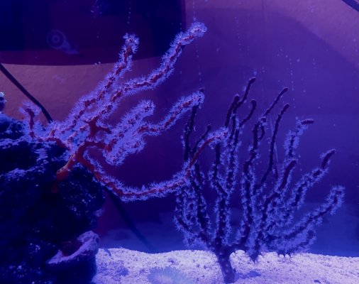
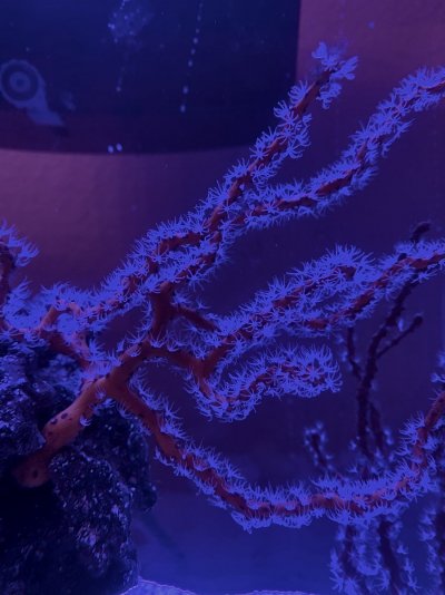
• Red Branching Gorgonian coral
• 1 encrusting montipora frag
• Purple micro-goniopora frag
Treatment Group consisted of:
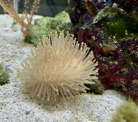
• 2 ricordea frags (one bleached, one healthy)
• 1 metallic neon green rhodactus
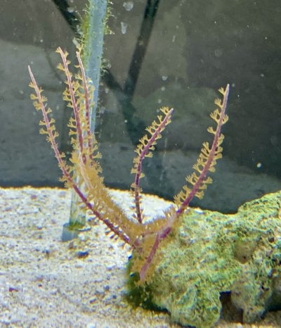
• Yellow Branching Gorgonian coral
• 1 encrusting montipora frag
• Peach micro-goniopora frag.
Materials:
• line, pump, and air stone
• 100% pure clove oil extract (which means no ethanol is added)
• display marine tank water (for rinsing)
• sterile saltwater (5 gallons)
• timer
• waste bucket
Procedures:
Calculated the dosage of clove oil extract to mix with the 2 gallons of display tank water to achieve a level of 1500ppm. Heavily aerated the tub for 30mins before bathing the treatment coral group. Started the 10min timer, and rinsed off each frag multiple times (3-5 rinses) with aged and clean saltwater at the end of the 10min duration.
After the bath and rinse, I took 24hr daily photos of the corals to determine if the results could be replicated (i.e. measuring polyp extensions) and measured the length of the polyps by using the iPhone measurement app.
Link to the academic study of clove oil as a potential drug option to treat cilliate infection in goniopora sp.: https://mdpi-res.com/d_attachment/biology/biology-11-00280/article_deploy/biology-11-00280.pdf
Results and Discussion:
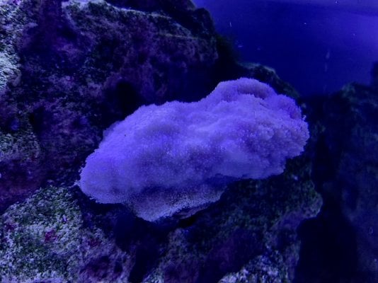
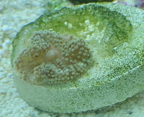
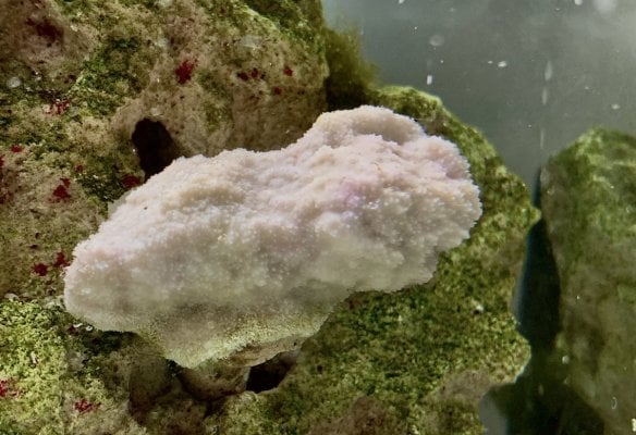
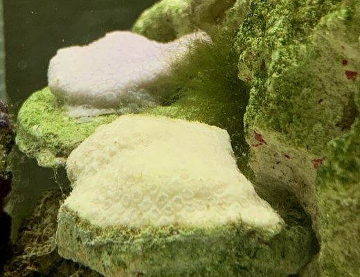
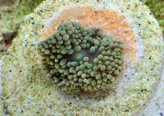
At the end of the initial 24 hours, a significant difference was found when comparing the Control Group with the Treatment Group; polyp extensions were shorter in the Control group while the Treatment group was visually healthier. A week later (today), here are the coral’s pictures after the clove oil bath. The bleached ricordea is growing back, as shown in the attachments.


