Internal Fish Diseases

Jay Hemdal

10K Club member
View Badges
Joined
Jul 31, 2020
Messages
28,604
Reaction score
28,261
Location
Dundee, MI
Rating - 0%
0   0   0
Internal Diseases

Cause(s)
A variety of organisms can infect fish internally. Most are worms, a few are protozoans, and there is even an eel that infects the heart of living sharks! Some species have complicated life cycles with intermediate hosts, and others can directly infect fish.

Nematodes can infect the gut of fish, causing very serious infections. In severe infections, they can migrate out of the gut and attack other organs. Tapeworms (cestodes) may also be present but do not reproduce without a second host, so they are rarely a problem. Gut protozoans, such as Hexamita, are normally present in most fish but sometimes cause disease when their populations grow as a result of the fish’s immune response being limited by some stress factor.

Hexamita was once implicated in causing head and lateral line erosion, but its presence in the guts of fish with HLLE was purely coincidental. If non-infected fish had been examined, Hexamita would have been found in them as well.

Sporozoans, such as myxosporidians, can internally infect fish. Myxobolus cerebralis is a parasite of salmonids (salmon and trout) that causes whirling disease. At least two species of myxosporidian fish parasites require a worm as an intermediary host. Henneguya salminicola is another species that infects salmonids. Infected fish have numerous large white cysts (up to 1 cm) in their skeletal muscle. When ruptured, the cysts release a white liquid filled with tiny myxozoan spores (~10 µm). No doubt, these parasites are well-known in salmonids due to their economic impact. It is presumed that other fish have similar myxosporidian infections but are just less studied. There is no treatment for myxosporian infections, but luckily, they are a relatively rare affliction in aquarium fishes.

Microsporidians were once thought to be protozoans but are now considered more closely related to fungi. They are obligate intracellular parasites, and create lesions called xenomas. One of the more commonly seen, called Glugea, creates white, smooth masses inside and outside of infected fish. Some people may mistake these growths for Cryptocaryon, but any spot that stays in the same position on a fish for more than a few days is not Cryptocaryon. Pseudoloma neurophilia is a common microsporidian pathogen found in zebrafish (Danio rerio) in research facilities. It causes emaciation and skeletal deformities, and thus may be confused with the symptoms of Mycobacterium sp. infections. There is no routine treatment for these diseases but borrowing from a treatment used to control Nosema infections in honeybees, one experimental treatment has been proposed: Fumagillin DCH administered in food, at 0.1 g/kg food at 1.5% body weight daily ration for four weeks. This seemed to prevent mortalities in one report.

One commonly seen microsporidian in tropical freshwater aquariums is “neon tetra disease” Pleistophora hyphessobryconis. Infecting not only neons, but other tetras and cyprinids, it is not directly treatable. Ironically, the similar-looking cardinal tetra, Paracheirodon axelrodi, appears to be mostly immune to this disease. Symptoms vary, but include whitening of the muscles of the fish, especially near the caudal peduncle, as well as white slimy patches on the skin (which can mimic Columnaris disease). Other symptoms can include overall “tattered” look, emaciation and pale coloration. Pleistophora is thought to transfer between fish when dead fish are cannibalized by others. Keeping the aquarium clean and promptly removing any dead fish may help control the spread of this disease.

Heterosporis sp. is another group of microsporidians. They occur within the skeletal muscle cells of fish, where it creates sporophorocysts up to 200 µm in diameter. New spores, 7-10 µm across grow inside vesicles found inside the sporophorocysts. Muscle tissue turns opaque and cloudy in affected fish, sometimes appearing granular. In gamefish, this makes the flesh unappetizing to anglers. This microsporidian group has been isolated from a wide range of freshwater fishes, including many aquarium species; angelfish (Pterophyllum scalare), Betta (Betta splendens), Loricariid catfish and Cichlids. Currently, there is no known treatment, and the level of morbidity seen in various fish is unknown. If a necropsy is not performed, infections will not be identified. Even with a necropsy, focus is generally made on internal organs, skin and gills, so sporophorocysts in the muscles can easily be missed.


Symptoms
Symptoms of internal parasites, like the causative organisms themselves, are varied. The most important visual symptom is emaciation—abnormal thinness of the fish’s belly and the nape (behind the head). If this is present and the fish is feeding well, then internal parasites of the gut may be competing for that food with the fish. Other symptoms include white, stringy feces and bloating of the abdomen.

Diagnosis
Internal parasites are extremely difficult to diagnose visually based on symptoms alone. As with bacterial infections (which are too small to see without high powered microscopes and cellular staining), these internal infections are often attributed to any problem in a fish that involves mucus being excreted with feces. Also, abnormal thinness can often be attributed to the fish not feeding well in the first place—it may be a species that does not feed well on normal aquarium fare and has no internal worms at all.

Looking at a fecal sample under a microscope may help with diagnosis but understand that any fecal matter deposited in an aquarium will become contaminated with all sorts of free-living protozoans and metazoans in a matter of an hour or so. If possible, collect a fish’s feces while it is being held in a clean container.

Treatment options
Wait out “self-limiting” parasites
As alluded to above, many internal parasites require additional hosts to complete their life cycle. In aquariums, these hosts are almost never present, so the disease is considered “self-limiting.” That is, once the adult parasite dies, the fish is free of the disease because no new parasites are being produced. In other
words, the “parasite load” never gets any higher than when the fish arrived in your aquarium. In such cases (like tapeworm infections), it is safest to just wait out the infection.

Some internal parasites have direct development; they do not need a secondary host and may not even need to leave the fish in order to complete their life cycle. These parasites, such as some nematodes, can become very serious infections and must be treated if possible.

Medicated foods
Medicated foods would seem to be the favored treatment option, but all are dosed as a percent of medication in the food, fed to the fish at a rate that is a proportion of the fish’s body weight. The trouble, of course, is that few aquarists know the weight of their fish.

Avoid the advice to soak the fish’s food with medication. This can never work except by pure luck. Think about it; nobody knows how much of the medication actually soaks into the food (if any), and then, since you don’t know the weight of the fish, the amount of food to feed is just a guess.

Garlic
One treatment that has found much favor with home aquarists is the use of garlic as a food additive. It is relatively non-toxic, so dosage errors are not dangerous to the fish. However, it is more of an “irritant” to internal parasites in the gut, dislodging some, but not all of them. Additionally, it does not seem to have any effect on internal parasites that have migrated out of the gut (such as some nematode worms). Its use
should be considered an adjunct therapy as opposed to a medical treatment. The amount of garlic to be used is not well defined, but there are commercial products available to use for soaking food. In some cases, people have over-extrapolated the benefit of garlic and use it to treat external parasites. There is little evidence that garlic as a food additive can control acute external parasitic infections.

Medicating the water
Since, by definition, these parasites are internal, external bath medications won’t easily reach them. However, marine fish do “drink” water in order to help maintain a proper osmotic balance, so medications added to the water itself do end up inside the fish—but at an unknown dosage. A typical marine teleost will drink on the order of 10–20% of their body weight per day, with the ability to drink up to 35–40% if the salinity is high. For example, a fish weighing 500 g will consume around 100 mL of saltwater in a day. It could then be possible to calculate how much medication is swallowed by a fish in 24 hours.

There is some anecdotal evidence that Praziquantel, dosed at 2.2 to 4 mg/l, can help eliminate internal parasites in marine fish. Dosing the water of a freshwater aquarium will not work to treat internal diseases of those fish as they do not ingest water, so none of the medication would be taken it via that route. Medicated food is a better choice for treating freshwater fishes for gut parasites.
 

Sharkbait19

10K Club member
View Badges
Joined
Aug 13, 2020
Messages
11,279
Reaction score
13,855
Location
New Jersey
Rating - 0%
0   0   0
Thanks for the info Jay! Great write up!
With fw fish, since meds can’t be directly dosed to the water, if they are at the point of not eating, is there anything that can be done?
 
OP
OP
Jay Hemdal

Jay Hemdal

10K Club member
View Badges
Joined
Jul 31, 2020
Messages
28,604
Reaction score
28,261
Location
Dundee, MI
Rating - 0%
0   0   0
Thanks for the info Jay! Great write up!
With fw fish, since meds can’t be directly dosed to the water, if they are at the point of not eating, is there anything that can be done?
Injectable meds is one option, but the best thing would be to tube feed liquid food with the proper treatment added. You need a fish anesthetic feeding tube and an accurate gram scale, to do that though.
 

LAReefer4Life

Lover of Angelfish
View Badges
Joined
Feb 11, 2019
Messages
2,989
Reaction score
8,265
Location
LOS ANGELES CA
Rating - 0%
0   0   0
After reading this, my takeaway from the article is there is not any effective treatments against internal disease??

How are you supposed to treat the fish effectively if medicated food nor treating the water column doesn't work? :thinking-face:
 
OP
OP
Jay Hemdal

Jay Hemdal

10K Club member
View Badges
Joined
Jul 31, 2020
Messages
28,604
Reaction score
28,261
Location
Dundee, MI
Rating - 0%
0   0   0
After reading this, my takeaway from the article is there is not any effective treatments against internal disease??

How are you supposed to treat the fish effectively if medicated food nor treating the water column doesn't work? :thinking-face:

For marine fish, dosing the water works in many cases because the fish drink the water. That doesn't work for freshwater fish.

Medicated food works great, but you just can't "soak food in medicine" like so many people do - it needs to be dosed properly:

Tube-feeding also works to deliver medication in fish that aren't feeding:

Jay
 

BubblesandSqueak

Valuable Member
View Badges
Joined
May 8, 2023
Messages
1,739
Reaction score
3,120
Location
Maine
Rating - 0%
0   0   0
Tube-feeding also works to deliver medication in fish that aren't feeding:

Jay
I'm assuming tube feeding is only for large fish? That can't be an easy task.
 
OP
OP
Jay Hemdal

Jay Hemdal

10K Club member
View Badges
Joined
Jul 31, 2020
Messages
28,604
Reaction score
28,261
Location
Dundee, MI
Rating - 0%
0   0   0
I'm assuming tube feeding is only for large fish? That can't be an easy task.

You need to have a fish anesthetic. I've tubed fish down to about 3 to 4" long using a tom cat catheter. I have also ruptured the stomachs of small fish trying to place the tube.

Jay
 

IrezumiHurts

Community Member
View Badges
Joined
Feb 13, 2023
Messages
83
Reaction score
107
Location
Seattle
Rating - 0%
0   0   0
@Jay Hemdal , I think it would be nice to see uronema added to this writeup as well, especially the challenges associated dealing with internal or intracellular afflictions.
 
OP
OP
Jay Hemdal

Jay Hemdal

10K Club member
View Badges
Joined
Jul 31, 2020
Messages
28,604
Reaction score
28,261
Location
Dundee, MI
Rating - 0%
0   0   0
@Jay Hemdal , I think it would be nice to see uronema added to this writeup as well, especially the challenges associated dealing with internal or intracellular afflictions.

I've never been able to successfully treat internal Uronema. I may have prevented its spread in some cases using chloroquine, but once a fish shows true Uronema lesions, the damage is already done.

Here is an older article I had written on Uronema:


IrezumiHurts

Community Member
View Badges
Joined
Feb 13, 2023
Messages
83
Reaction score
107
Location
Seattle
Rating - 0%
0   0   0
I've never been able to successfully treat internal Uronema. I may have prevented its spread in some cases using chloroquine, but once a fish shows true Uronema lesions, the damage is already done.

Here is an older article I had written on Uronema:

@Jay Hemdal I'm curious to know your thoughts on this then, I have a Regal angel that made it 29 days through comprehensive QT minus formalin. Day 29, presented with initial signs of uronema - both lateral bands and circular sores, lifting scales red sores and all. You know the look. Quite a few small pink spots that I would call "pre-fissure" as well. I've thrown the kitchen sink at him and somehow seem to arrested the progression of the disease (but not cured it). Would this imply that im likely dealing with an external uronema affliction, or just somehow delaying the inevitable? Details to follow if interested.

While I certainly don't have your level of experience, I have become pretty good at recognizing this disease recently so i don't think this is misdiagnosed. This 100g QT tank has claimed about 20 fish over the last 3 months. At first I blamed supply chain/cyanide, but then when putting a batch of angels and butterflies through, it claimed almost every single one of them - half presented with the classic red sores, it was immediately evident what I was dealing with. I had suspicions early on (but had yet to knowingly encounter uronema to this point) so i purchased formalin and sent for Aquabiomics eDNA tests, which came back with positive confirmation on marinum. Unfortunately the results were too late, and came while I already had this Regal in QT mid cycle.

Fast forward to 7 days ago, once the signs emerged (the ones listed above) i immediately kicked into gear. I know formalin is not advised once sores appears due to the aggressive nature of the chemical, but my estimation they were light enough i could attempt treatment with formalin without causing too much undue pain and stress to the fish.

Regal was immediately transferred to a heavily aerated bath, where I ramped the formalin every 5 minutes (for pain management) to a final target of 1ml/gal over 30 minutes, and held at full strength for 1 hour. I am aware it is recommended to treat at 0.6ml/gal for temps over 70F, but I felt I had nothing to lose being aggressive.

I also did a 100%WC and dosed the QT tank 1ml/10gal every 24 hours and added extra filtration. Original non- absorbing biomedia was reused to manage ammonia under the pretense the in- tank formalin levels would eliminate it.

I have continued bath treatments on the angel every 3 days now, as well a adding 2 additional baths - Ruby rally 90 minute bath before the 1hr formalin bath, and 1hr nitrofurizone/kanamyacin/methylene blue bath after the formalin, before transfer back into the formalin-dosed QT. I also started adding melafix at the in-tank dosage prescribed on the bottle. Again, kitchen sink, I'm throwing everything I can at him, even if it's experimental at best.

Previous encounters with this disease had mortality occurring within about 72 hours from initial visible ruptures at the skin surface or when I could see lifting of scales. The sores would also rapidly multiply and increase in both intensity and the redness.

This far the symptoms have not cleared, but quite literally have arrested in progress, and some of the redness has gone down or disappeared. I don't know what to make of this, I was not anticipating any degree of success.

I am concerned about continued use of formalin however, I'm currently on day 8, and was considering switching from the current treatment to chloroquine phosphate (I have some on hand, properly stored) at the higher 80mg dosage level. What would you do in this circumstance? Do uronema sores always = internal uronema?
 
OP
OP
Jay Hemdal

Jay Hemdal

10K Club member
View Badges
Joined
Jul 31, 2020
Messages
28,604
Reaction score
28,261
Location
Dundee, MI
Rating - 0%
0   0   0
@Jay Hemdal I'm curious to know your thoughts on this then, I have a Regal angel that made it 29 days through comprehensive QT minus formalin. Day 29, presented with initial signs of uronema - both lateral bands and circular sores, lifting scales red sores and all. You know the look. Quite a few small pink spots that I would call "pre-fissure" as well. I've thrown the kitchen sink at him and somehow seem to arrested the progression of the disease (but not cured it). Would this imply that im likely dealing with an external uronema affliction, or just somehow delaying the inevitable? Details to follow if interested.

While I certainly don't have your level of experience, I have become pretty good at recognizing this disease recently so i don't think this is misdiagnosed. This 100g QT tank has claimed about 20 fish over the last 3 months. At first I blamed supply chain/cyanide, but then when putting a batch of angels and butterflies through, it claimed almost every single one of them - half presented with the classic red sores, it was immediately evident what I was dealing with. I had suspicions early on (but had yet to knowingly encounter uronema to this point) so i purchased formalin and sent for Aquabiomics eDNA tests, which came back with positive confirmation on marinum. Unfortunately the results were too late, and came while I already had this Regal in QT mid cycle.

Fast forward to 7 days ago, once the signs emerged (the ones listed above) i immediately kicked into gear. I know formalin is not advised once sores appears due to the aggressive nature of the chemical, but my estimation they were light enough i could attempt treatment with formalin without causing too much undue pain and stress to the fish.

Regal was immediately transferred to a heavily aerated bath, where I ramped the formalin every 5 minutes (for pain management) to a final target of 1ml/gal over 30 minutes, and held at full strength for 1 hour. I am aware it is recommended to treat at 0.6ml/gal for temps over 70F, but I felt I had nothing to lose being aggressive.

I also did a 100%WC and dosed the QT tank 1ml/10gal every 24 hours and added extra filtration. Original non- absorbing biomedia was reused to manage ammonia under the pretense the in- tank formalin levels would eliminate it.

I have continued bath treatments on the angel every 3 days now, as well a adding 2 additional baths - Ruby rally 90 minute bath before the 1hr formalin bath, and 1hr nitrofurizone/kanamyacin/methylene blue bath after the formalin, before transfer back into the formalin-dosed QT. I also started adding melafix at the in-tank dosage prescribed on the bottle. Again, kitchen sink, I'm throwing everything I can at him, even if it's experimental at best.

Previous encounters with this disease had mortality occurring within about 72 hours from initial visible ruptures at the skin surface or when I could see lifting of scales. The sores would also rapidly multiply and increase in both intensity and the redness.

This far the symptoms have not cleared, but quite literally have arrested in progress, and some of the redness has gone down or disappeared. I don't know what to make of this, I was not anticipating any degree of success.

I am concerned about continued use of formalin however, I'm currently on day 8, and was considering switching from the current treatment to chloroquine phosphate (I have some on hand, properly stored) at the higher 80mg dosage level. What would you do in this circumstance? Do uronema sores always = internal uronema?

I've only seen external Uronema in seahorses and sea dragons, it may even be a different genus (Miamiensis or Philasterides)..

The issue with Aquabiomics and Uronema is that this is a facultative parasite, its normal "day job" is feeding on bacteria. Estimates range from 25 to 75% of aquariums have a population of Uronema in residence, with no overt sign of disease in the fish. Nobody knows how the internal Uronema gets inside the fish, but it seems to happen during the fish's trip through the supply chain.

1 ml of formalin per gallon works out to be over 250 ppm, that is too high for tropical fish, even with good aeration. If you are doing one hour dips, you should never go higher than 167 ppm, I use 150 ppm. It isn't just the formalin, it is the methanol used as a preservative, as well as the oxygen reduction from the formalin. In addition, you've got to be REALLY careful using that in a home.

I've never heard of ramping up formalin dips, where did you hear of that? The trouble is that formalin is dosed on time versus concentration, and having a sliding concentration would mess with the dose too much to know where you are at.

The trouble is that Uronema is a great imitator of other issues, mostly bacterial. In fact, Uronema can co-habitat with bacteria (which they feed on). To be 100% certain you are dealing with uronema, you need to do a skin biopsy/wet mount. Of course, that's pretty rough on the fish.

What is that 80 mg chloroquine dose? 80 mg / gallon? That's over 20 mg/l, I started running into toxicity issues in that range.
 

IrezumiHurts

Community Member
View Badges
Joined
Feb 13, 2023
Messages
83
Reaction score
107
Location
Seattle
Rating - 0%
0   0   0
I've only seen external Uronema in seahorses and sea dragons, it may even be a different genus (Miamiensis or Philasterides)..

The issue with Aquabiomics and Uronema is that this is a facultative parasite, its normal "day job" is feeding on bacteria. Estimates range from 25 to 75% of aquariums have a population of Uronema in residence, with no overt sign of disease in the fish. Nobody knows how the internal Uronema gets inside the fish, but it seems to happen during the fish's trip through the supply chain.

1 ml of formalin per gallon works out to be over 250 ppm, that is too high for tropical fish, even with good aeration. If you are doing one hour dips, you should never go higher than 167 ppm, I use 150 ppm. It isn't just the formalin, it is the methanol used as a preservative, as well as the oxygen reduction from the formalin. In addition, you've got to be REALLY careful using that in a home.

I've never heard of ramping up formalin dips, where did you hear of that? The trouble is that formalin is dosed on time versus concentration, and having a sliding concentration would mess with the dose too much to know where you are at.

The trouble is that Uronema is a great imitator of other issues, mostly bacterial. In fact, Uronema can co-habitat with bacteria (which they feed on). To be 100% certain you are dealing with uronema, you need to do a skin biopsy/wet mount. Of course, that's pretty rough on the fish.

What is that 80 mg chloroquine dose? 80 mg / gallon? That's over 20 mg/l, I started running into toxicity issues in that range.

@Jay Hemdal Thanks again for the responses.

Yes to be clear I am using PPE (3M 6005 cartridges) , gloves goggles and outdoors in a ventilated makeshift greenhouse. I read about the issues with this chemical and I know you personally had issues I read somewhere when you were 14yrs old working in a fish farm the formalin would burn your eyes. That's really terrible so thank you for advocating safety.

I am tracking on the issue of uronema preferring bacteria etc. In fact I did tests on all of my tanks - one of my DTs had a strain called "heteromarinum" but I've never had an issue in it for about 2 years now. This tank also has 2x chromis, 1x borblnius and 2x lyretail anthias, and a flame angel since the beginning. Actually I've had 0 fish deaths in this tank aside from 2x BSJs jumping. The only tank that I've had issues in was my large QT, which tested positive for marinum. So either heteromarinum doesn't prefer fish, or there is enough bioavailable food otherwise. Incidentally that DT tank went for 6 months with no filtration and no water changes, 600ppm Nitrates, so there certainly wasn't any cleanliness to inhibit proliferation of the heteromarinum in that tank. I cleaned an inch thick orange mat of detritus from the sump. Yes you read that right, it's a long story but the short story is the FOWLR tank was under someone else's care. The fish and inverts seemed to have cared less. I digress.

The reason for ramping the formalin, was my fear the formalin burns an open wound, maybe like rubbing alcohol, and I didn't want to induce unnecessary stress. My unscientific reasoning was by ramping the medication, it would ease the fish into the process. From a stress standpoint it seemed to work though it was spittng water at the surface and swimming into the bubbles at the 57 minute mark. I promptly pulled him.

Yes I am aware this is a very high/ above recommended formalin dose, an yes I am running it for an hour but as I mentioned it doesn't reach target strength until 30 minutes. My future dips planned to be at lower strength (0.6ml/gal) without the ramp due to the apparent efficacy/stabilization of the treatments thus far.

You read correctly on my intent to transition to 80mg/gal CP, if I choose to migrate away from the formalin treatment. Though along the reasoning in my above paragraph, would likely consider dropping to 60 or 70mg/gal, since the situation appears to be stabilizing. I understand 80mg/gal is at the extreme upper end of what's tolerated by fish - to the extent that many cannot. My rationale being I am going to lose this fish to uronema anyways, I should at least try until I see the symptoms devolve to a point where I need to euthanize. I have not reached that point.

Regarding a skin scrape I do not have a microscope and this may sound ignorant but truthfully I would probably not scrape even if I did in this circumstance, to avoid additional stress or damage to the fish and also because I am quite confident in my assessment of the uronema issues against the context of what I've endured the past few months, paired with the days from the eDNA test. I know that's not scientific per say (one issue doesn't always beget the other) but for me this is just an occams razor/ common sense thing. The circumstantial evidence would indicate I am dealing with a bad urinema marinum issue in my QT tank which has aggressively killed multiple fish over the last few months, and so far aside from the treatment actually seeming to be stabilization the fish, everything seems to support it being uronema. I will attach a few pictures of a few previous mortalities as well a the Regal. Pictures with the yellow figure font were from 8 days ago, unlabeled ones were yesterday. Sorry I didn't mean to hijack this thread so if this needs to be moved elsewhere that's fine too.
 

IrezumiHurts

Community Member
View Badges
Joined
Feb 13, 2023
Messages
83
Reaction score
107
Location
Seattle
Rating - 0%
0   0   0
Try again
20240117_192645.jpg
20240115_092126.jpg
20240512_015704.jpg
20240512_015652.jpg
20240512_015717.jpg
20240512_015726.jpg
20240516_195141.jpg
20240516_195206.jpg
 
OP
OP
Jay Hemdal

Jay Hemdal

10K Club member
View Badges
Joined
Jul 31, 2020
Messages
28,604
Reaction score
28,261
Location
Dundee, MI
Rating - 0%
0   0   0
Try again
20240117_192645.jpg
20240115_092126.jpg
20240512_015704.jpg
20240512_015652.jpg
20240512_015717.jpg
20240512_015726.jpg
20240516_195141.jpg
20240516_195206.jpg

Just based on gross visual symptoms, the Heniochus has classic Uronema. The Navarchus angel is odd because the lesions look broader and shallower, and seeing two of them on the same fish, and having the fish still be up and swimming is not typical. The regal seems to have multiple small spot, that also isn't typical of Uronema.

Still, acute, contagious bacterial infections are also very rare.

You can get a microscope for your cell phone for about $30. It can be used on dead fish (if they are fresh) to identify protozoans. They won't visualize bacteria though.
 

IrezumiHurts

Community Member
View Badges
Joined
Feb 13, 2023
Messages
83
Reaction score
107
Location
Seattle
Rating - 0%
0   0   0
Just based on gross visual symptoms, the Heniochus has classic Uronema. The Navarchus angel is odd because the lesions look broader and shallower, and seeing two of them on the same fish, and having the fish still be up and swimming is not typical. The regal seems to have multiple small spot, that also isn't typical of Uronema.

Still, acute, contagious bacterial infections are also very rare.

You can get a microscope for your cell phone for about $30. It can be used on dead fish (if they are fresh) to identify protozoans. They won't visualize bacteria though.
The majestic started out with the same lesions as the heniochus but this photo was terminal stage - he did not survive more than a few hours after this photo. He was patient 0 so I wish I had done better at documenting the symptoms progression, my first thought was a tang had sliced his sides because there was multiple thin seams along his side that would open up when he flexed the opposite direction where i could see a bright red line under the lifting scales. Of course this was a silly assumption in retrospect. I placed him into a ciprofloxacin hospital tank which did nothing. These thin seams soon became red and enlarged and finally over the course of 24 hrs the very broad sores you see at the end. He had become incredibly reclusive and stopped eating a few days leading up to the initial symptoms, more reclusive than these fish typically are anyways.

I'll look into the microscope for the phone, never knew such a thing existed. If he passes I'll document the necropsy. I think in the meantime I'm going to transition from formalin to CP and hope for the best.
 
OP
OP
Jay Hemdal

Jay Hemdal

10K Club member
View Badges
Joined
Jul 31, 2020
Messages
28,604
Reaction score
28,261
Location
Dundee, MI
Rating - 0%
0   0   0
Here is a really cheap cell phone microscope that I bought:

60x Zoom Microscope Magnifier LED + Uv Light Clip-on Micro Lens for Universal Mobile Phones Universal​

Here is a better one:

APEXEL Phone Macro Lens, 100X Microscope for Android/iPhone Micro Camera with LED​

 
Last edited:

Hossein

New Member
View Badges
Joined
May 30, 2024
Messages
2
Reaction score
0
Location
Iran
Rating - 0%
0   0   0
Internal Diseases

Cause(s)
A variety of organisms can infect fish internally. Most are worms, a few are protozoans, and there is even an eel that infects the heart of living sharks! Some species have complicated life cycles with intermediate hosts, and others can directly infect fish.

Nematodes can infect the gut of fish, causing very serious infections. In severe infections, they can migrate out of the gut and attack other organs. Tapeworms (cestodes) may also be present but do not reproduce without a second host, so they are rarely a problem. Gut protozoans, such as Hexamita, are normally present in most fish but sometimes cause disease when their populations grow as a result of the fish’s immune response being limited by some stress factor.

Hexamita was once implicated in causing head and lateral line erosion, but its presence in the guts of fish with HLLE was purely coincidental. If non-infected fish had been examined, Hexamita would have been found in them as well.

Sporozoans, such as myxosporidians, can internally infect fish. Myxobolus cerebralis is a parasite of salmonids (salmon and trout) that causes whirling disease. At least two species of myxosporidian fish parasites require a worm as an intermediary host. Henneguya salminicola is another species that infects salmonids. Infected fish have numerous large white cysts (up to 1 cm) in their skeletal muscle. When ruptured, the cysts release a white liquid filled with tiny myxozoan spores (~10 µm). No doubt, these parasites are well-known in salmonids due to their economic impact. It is presumed that other fish have similar myxosporidian infections but are just less studied. There is no treatment for myxosporian infections, but luckily, they are a relatively rare affliction in aquarium fishes.

Microsporidians were once thought to be protozoans but are now considered more closely related to fungi. They are obligate intracellular parasites, and create lesions called xenomas. One of the more commonly seen, called Glugea, creates white, smooth masses inside and outside of infected fish. Some people may mistake these growths for Cryptocaryon, but any spot that stays in the same position on a fish for more than a few days is not Cryptocaryon. Pseudoloma neurophilia is a common microsporidian pathogen found in zebrafish (Danio rerio) in research facilities. It causes emaciation and skeletal deformities, and thus may be confused with the symptoms of Mycobacterium sp. infections. There is no routine treatment for these diseases but borrowing from a treatment used to control Nosema infections in honeybees, one experimental treatment has been proposed: Fumagillin DCH administered in food, at 0.1 g/kg food at 1.5% body weight daily ration for four weeks. This seemed to prevent mortalities in one report.

One commonly seen microsporidian in tropical freshwater aquariums is “neon tetra disease” Pleistophora hyphessobryconis. Infecting not only neons, but other tetras and cyprinids, it is not directly treatable. Ironically, the similar-looking cardinal tetra, Paracheirodon axelrodi, appears to be mostly immune to this disease. Symptoms vary, but include whitening of the muscles of the fish, especially near the caudal peduncle, as well as white slimy patches on the skin (which can mimic Columnaris disease). Other symptoms can include overall “tattered” look, emaciation and pale coloration. Pleistophora is thought to transfer between fish when dead fish are cannibalized by others. Keeping the aquarium clean and promptly removing any dead fish may help control the spread of this disease.

Heterosporis sp. is another group of microsporidians. They occur within the skeletal muscle cells of fish, where it creates sporophorocysts up to 200 µm in diameter. New spores, 7-10 µm across grow inside vesicles found inside the sporophorocysts. Muscle tissue turns opaque and cloudy in affected fish, sometimes appearing granular. In gamefish, this makes the flesh unappetizing to anglers. This microsporidian group has been isolated from a wide range of freshwater fishes, including many aquarium species; angelfish (Pterophyllum scalare), Betta (Betta splendens), Loricariid catfish and Cichlids. Currently, there is no known treatment, and the level of morbidity seen in various fish is unknown. If a necropsy is not performed, infections will not be identified. Even with a necropsy, focus is generally made on internal organs, skin and gills, so sporophorocysts in the muscles can easily be missed.


Symptoms
Symptoms of internal parasites, like the causative organisms themselves, are varied. The most important visual symptom is emaciation—abnormal thinness of the fish’s belly and the nape (behind the head). If this is present and the fish is feeding well, then internal parasites of the gut may be competing for that food with the fish. Other symptoms include white, stringy feces and bloating of the abdomen.

Diagnosis
Internal parasites are extremely difficult to diagnose visually based on symptoms alone. As with bacterial infections (which are too small to see without high powered microscopes and cellular staining), these internal infections are often attributed to any problem in a fish that involves mucus being excreted with feces. Also, abnormal thinness can often be attributed to the fish not feeding well in the first place—it may be a species that does not feed well on normal aquarium fare and has no internal worms at all.

Looking at a fecal sample under a microscope may help with diagnosis but understand that any fecal matter deposited in an aquarium will become contaminated with all sorts of free-living protozoans and metazoans in a matter of an hour or so. If possible, collect a fish’s feces while it is being held in a clean container.

Treatment options
Wait out “self-limiting” parasites
As alluded to above, many internal parasites require additional hosts to complete their life cycle. In aquariums, these hosts are almost never present, so the disease is considered “self-limiting.” That is, once the adult parasite dies, the fish is free of the disease because no new parasites are being produced. In other
words, the “parasite load” never gets any higher than when the fish arrived in your aquarium. In such cases (like tapeworm infections), it is safest to just wait out the infection.

Some internal parasites have direct development; they do not need a secondary host and may not even need to leave the fish in order to complete their life cycle. These parasites, such as some nematodes, can become very serious infections and must be treated if possible.

Medicated foods
Medicated foods would seem to be the favored treatment option, but all are dosed as a percent of medication in the food, fed to the fish at a rate that is a proportion of the fish’s body weight. The trouble, of course, is that few aquarists know the weight of their fish.

Avoid the advice to soak the fish’s food with medication. This can never work except by pure luck. Think about it; nobody knows how much of the medication actually soaks into the food (if any), and then, since you don’t know the weight of the fish, the amount of food to feed is just a guess.

Garlic
One treatment that has found much favor with home aquarists is the use of garlic as a food additive. It is relatively non-toxic, so dosage errors are not dangerous to the fish. However, it is more of an “irritant” to internal parasites in the gut, dislodging some, but not all of them. Additionally, it does not seem to have any effect on internal parasites that have migrated out of the gut (such as some nematode worms). Its use
should be considered an adjunct therapy as opposed to a medical treatment. The amount of garlic to be used is not well defined, but there are commercial products available to use for soaking food. In some cases, people have over-extrapolated the benefit of garlic and use it to treat external parasites. There is little evidence that garlic as a food additive can control acute external parasitic infections.

Medicating the water
Since, by definition, these parasites are internal, external bath medications won’t easily reach them. However, marine fish do “drink” water in order to help maintain a proper osmotic balance, so medications added to the water itself do end up inside the fish—but at an unknown dosage. A typical marine teleost will drink on the order of 10–20% of their body weight per day, with the ability to drink up to 35–40% if the salinity is high. For example, a fish weighing 500 g will consume around 100 mL of saltwater in a day. It could then be possible to calculate how much medication is swallowed by a fish in 24 hours.

There is some anecdotal evidence that Praziquantel, dosed at 2.2 to 4 mg/l, can help eliminate internal parasites in marine fish. Dosing the water of a freshwater aquarium will not work to treat internal diseases of those fish as they do not ingest water, so none of the medication would be taken it via that route. Medicated food is a better choice for treating freshwater fishes for gut parasites.
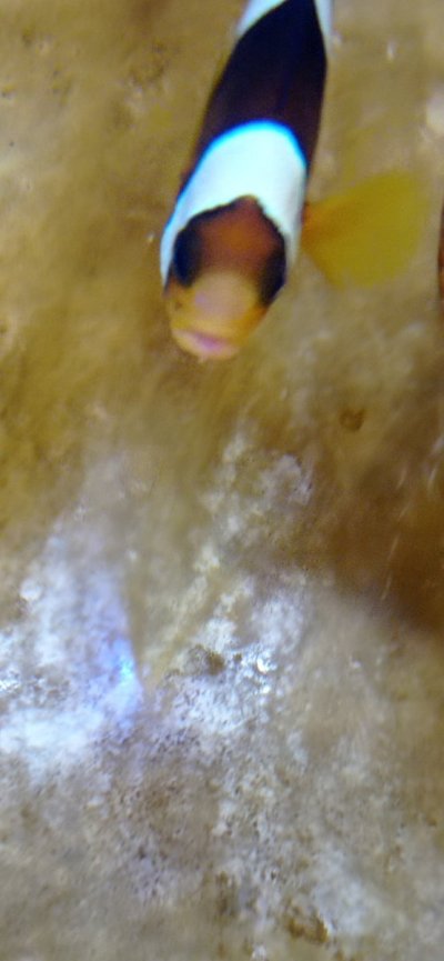
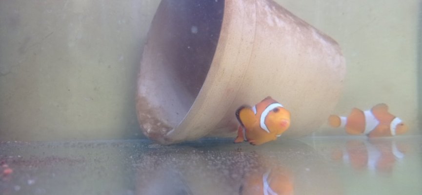
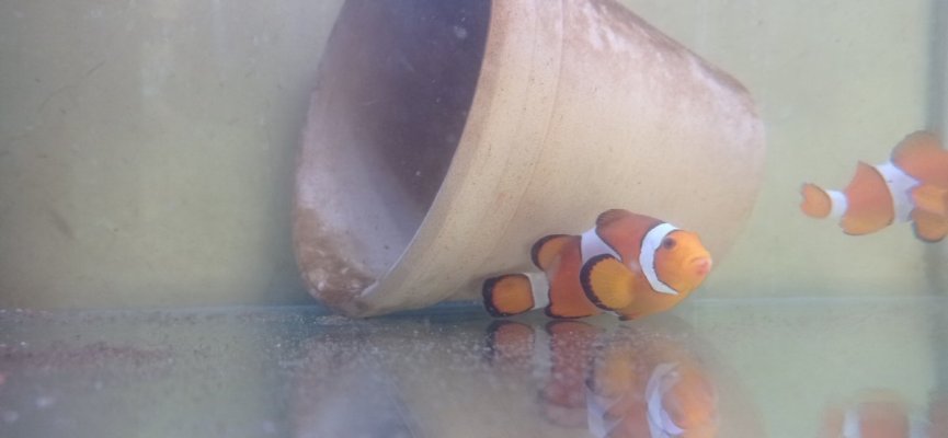
Dear Dr Jay Hemdal
In the attachment, the pictures of the sick clownfish with the signs of reddening of the mouth and the appearance of cotton on the lips of the fish on a very small scale.
The fish does not eat at all.
The red spots created under the lower jaw can be seen in both females and males.
Out of the 5 clown fish involved, 3 male fish and 2 female fish were involved.
And it doesn't seem to be caused by cleaning activities for spawning.
For example, in a breeding pair of Ocellaris clownfish
, only the female fish is involved and the male fish has no symptoms.
A red dot forms under the lower jaw and continues to progress and becomes an extra tissue under the jaw and inside the mouth.
So far, this disease has occurred in seven clown fish, five of which have died, and it affects exactly one area, that is, under the lower jaw.
Thank you very much for giving me your valuable time.
 

Attachments

  • IMG_20240514_063409.jpg
    IMG_20240514_063409.jpg
    81 KB · Views: 42
  • IMG_20240524_120446.jpg
    IMG_20240524_120446.jpg
    57.4 KB · Views: 39
  • IMG_20240524_120449.jpg
    IMG_20240524_120449.jpg
    58.4 KB · Views: 42
OP
OP
Jay Hemdal

Jay Hemdal

10K Club member
View Badges
Joined
Jul 31, 2020
Messages
28,604
Reaction score
28,261
Location
Dundee, MI
Rating - 0%
0   0   0
Dear Dr Jay Hemdal
In the attachment, the pictures of the sick clownfish with the signs of reddening of the mouth and the appearance of cotton on the lips of the fish on a very small scale.
The fish does not eat at all.
The red spots created under the lower jaw can be seen in both females and males.
Out of the 5 clown fish involved, 3 male fish and 2 female fish were involved.
And it doesn't seem to be caused by cleaning activities for spawning.
For example, in a breeding pair of Ocellaris clownfish
, only the female fish is involved and the male fish has no symptoms.
A red dot forms under the lower jaw and continues to progress and becomes an extra tissue under the jaw and inside the mouth.
So far, this disease has occurred in seven clown fish, five of which have died, and it affects exactly one area, that is, under the lower jaw.
Thank you very much for giving me your valuable time.
Lesions on the jaws of clownfish mouths are typically caused by “lip locking” - what clownfish do when establishing territory and dominance. However, seeing this in a number of clownfish that died is unusual. When clownfish fight, you may never see it because when you are in the room, they stop fighting to watch you. There is almost always a “winner” whose jaws are undamaged.

One other possibility is that this minor skin damage is allowing Mycobactetium marinum (fish tuberculosis) to enter the wound and start an infection. There is no good treatment for this however.
 

HAVE YOU EVER KEPT A RARE/UNCOMMON FISH, CORAL, OR INVERT? SHOW IT OFF IN THE THREAD!

  • Yes!

    Votes: 32 45.7%
  • Not yet, but I have one that I want to buy in mind!

    Votes: 9 12.9%
  • No.

    Votes: 26 37.1%
  • Other (please explain).

    Votes: 3 4.3%
Back
Top