Effect of UV-A Spectrum on Acropora Growth and Coloration

J1a

Well-Known Member
View Badges
Joined
Nov 14, 2021
Messages
666
Reaction score
950
Location
Singapore
Rating - 0%
0   0   0
Effect of UV-A Spectrum on Acropora Growth and Coloration

Hypothesis
:
  1. UV-A supplementation can increase the growth in acropora sp.
  2. UV-A supplementation can initate or increase the production of fluoroproteins in acropora sp.
Methodology :

Preparation :
  1. Two fresh frags from a single acropora are made and fixed on frag tiles with scale.
  2. The frags are labeled E (experimental) and C (control).
  3. The frags are placed in the same aquarium, approx 6" apart. A black acrylic sheet is placed in-between them.
  4. Using a spectrum photometer, measure the PAR and spectrum profile at both frags.
  5. Adjust the position of the frags to achieve matching PAR and spectrum.
  6. Culture the frag for one week to allow healing and observe encrustation.
  7. Test and record water parameter.
Intervention:
  1. Install a UV light strip (360nm, 380nm) over the experiment group.
  2. Ramp up the intensity of the UV over 1 week, to approx 15% of the original PAR.
Measurements:
  1. Diameter of encrusted base, weekly;
  2. Height of frag, weekly;
  3. Observable growth tips, weekly;
  4. The coloration will be measured by taking photos of both frags using manual camera mode, with identical exporsure parameters, and edited using identical process (saved preset in Adobe Photoshop Express), weekly;
Maybe there is something, maybe there is not. We shall see.

I do not have space in my house to do this. Therefore I will be collaborating with a LFS here in Singapore to carry out this investigation. Let me know if you have any suggestions to improve this process.

Thank you for your time!
 

taricha

5000 Club Member
View Badges
Joined
May 22, 2016
Messages
6,970
Reaction score
10,747
Rating - 0%
0   0   0
Ramp up the intensity of the UV over 1 week, to approx 15% of the original PAR.
I'd add a third group with the normal (control) spectrum, but increased the intensity to match the intensity of the UV-added. This would allow you to distinguish whether the changes observed with adding the UV are from the UV, or simply from the increased intensity.

The coloration will be measured by taking photos of both frags using manual camera mode, with identical exporsure parameters, and edited using identical process (saved preset in Adobe Photoshop Express), weekly;
To get this cleaner, data-wise, I'd pull the frags into the same space for the photographs, and shoot it with some sort of color reference card. Having the corals and the color reference in the same photo will make it pretty hard to fool yourself as to what's happening. You can map any significant RGB pixel shifts in the corals using the RGB of the color card as reference, and you can digitally adjust the photographs from one week to the next to make the pixel values of the color cards the same across the whole time period of the experiment.
Next best thing if you can't/don't want to move the corals for photo sessions, then you can situate the color card the same in photos of each and digitally adjust the photos to make the color cards match.
 
OP
OP
J1a

J1a

Well-Known Member
View Badges
Joined
Nov 14, 2021
Messages
666
Reaction score
950
Location
Singapore
Rating - 0%
0   0   0
I'd add a third group with the normal (control) spectrum, but increased the intensity to match the intensity of the UV-added. This would allow you to distinguish whether the changes observed with adding the UV are from the UV, or simply from the increased intensity.
I have contemplated about this. I was deciding between treating UV as addition or replacement. But yeah. I think can add on a C2 group for increased PAR based on the baseline spectrum.

To get this cleaner, data-wise, I'd pull the frags into the same space for the photographs, and shoot it with some sort of color reference card. Having the corals and the color reference in the same photo will make it pretty hard to fool yourself as to what's happening. You can map any significant RGB pixel shifts in the corals using the RGB of the color card as reference, and you can digitally adjust the photographs from one week to the next to make the pixel values of the color cards the same across the whole time period of the experiment.
Next best thing if you can't/don't want to move the corals for photo sessions, then you can situate the color card the same in photos of each and digitally adjust the photos to make the color cards match.

My original intention is to just capture RAW file with both frags in the same frame, they are pretty near to begin with. I would think using RAW and run the file through the same filter will probably eliminate any bias. But yeah. I can include a color reference card just to be sure.

Appreciate your inputs!
 

taricha

5000 Club Member
View Badges
Joined
May 22, 2016
Messages
6,970
Reaction score
10,747
Rating - 0%
0   0   0
My original intention is to just capture RAW file with both frags in the same frame, they are pretty near to begin with.
Oops, yeah. I missed the part that they are in the same tank with light dividers.

Digital cameras are way smarter than me. Color cards are the only way I know I can check their work. :)
 
OP
OP
J1a

J1a

Well-Known Member
View Badges
Joined
Nov 14, 2021
Messages
666
Reaction score
950
Location
Singapore
Rating - 0%
0   0   0
Oops, yeah. I missed the part that they are in the same tank with light dividers.

Digital cameras are way smarter than me. Color cards are the only way I know I can check their work. :)
I'll throw the cards in just in case something else comes up.
 

MnFish1

10K Club member
View Badges
Joined
Dec 28, 2016
Messages
24,326
Reaction score
23,111
Location
Midwest
Rating - 100%
1   0   0
Effect of UV-A Spectrum on Acropora Growth and Coloration

Hypothesis
:
  1. UV-A supplementation can increase the growth in acropora sp.
  2. UV-A supplementation can initate or increase the production of fluoroproteins in acropora sp.
Methodology :

Preparation :
  1. Two fresh frags from a single acropora are made and fixed on frag tiles with scale.
  2. The frags are labeled E (experimental) and C (control).
  3. The frags are placed in the same aquarium, approx 6" apart. A black acrylic sheet is placed in-between them.
  4. Using a spectrum photometer, measure the PAR and spectrum profile at both frags.
  5. Adjust the position of the frags to achieve matching PAR and spectrum.
  6. Culture the frag for one week to allow healing and observe encrustation.
  7. Test and record water parameter.
Intervention:
  1. Install a UV light strip (360nm, 380nm) over the experiment group.
  2. Ramp up the intensity of the UV over 1 week, to approx 15% of the original PAR.
Measurements:
  1. Diameter of encrusted base, weekly;
  2. Height of frag, weekly;
  3. Observable growth tips, weekly;
  4. The coloration will be measured by taking photos of both frags using manual camera mode, with identical exporsure parameters, and edited using identical process (saved preset in Adobe Photoshop Express), weekly;
Maybe there is something, maybe there is not. We shall see.

I do not have space in my house to do this. Therefore I will be collaborating with a LFS here in Singapore to carry out this investigation. Let me know if you have any suggestions to improve this process.

Thank you for your time!
Nice job.
 
OP
OP
J1a

J1a

Well-Known Member
View Badges
Joined
Nov 14, 2021
Messages
666
Reaction score
950
Location
Singapore
Rating - 0%
0   0   0
Background

One of the many goals of reef keeping is to achieve good growth and coloration of captive corals. Many corals, especially acroporidae, relies on their symbiotic zooxanthelle for nutrition budget. These corals also often exhibit fluorescence, presumably to modulate the recieved irradiation.

Therefore it is conceivable that the quanity and quality of light incident on the captive reef can have an impact on the growth and coloration of corals.


LED and Its Spectrum

The application of LED in reef lighting has seen huge improvement since its nascence. Many modern light sets are desgined with diodes matching the photosyntheic absorption of zooxanthelle. The blue heavy spectrum provide high PUR to PAR ratio, and at the same time, provide suitable excitation wavelengths to produce fluorescence in corals.

However, LED reef lights are largely unable to provide suitable UV-A spectrum, which many coral would recieve in the natural reef. Due to cost, efficiency and the demand on glass optics, most reef lights does not produce meaningful quanity of radiation in the 350-380nm region.

Will these spectrum affect the physiology of acropora? Is it worthwhile to supplement UV-A? These are the questions I will attempt to find answers to.


Anecdotal Evidence

My aquarium is brightly lit, with the top of my reef recieving 1.2k/(μmol/m2/s) PAR (actually, PPFD). The UV portion of the light is supplied by some 395nm diodes. There is no meaningful amount of radiation below 385nm.

In late December 2021, I installed a OR3 lightbar with 380nm and 420nm diodes to supplement the UV spectrum. The light bar produces negligible (<5%) PAR increase, but it is able to provide additional 30 PAR of radiation in the UV-A region. The light bar is turned on during the day light hours of my aquarium light set.

While no other changes were made to the aquarium husbandry, the difference is quite visible within one month of supplementation.

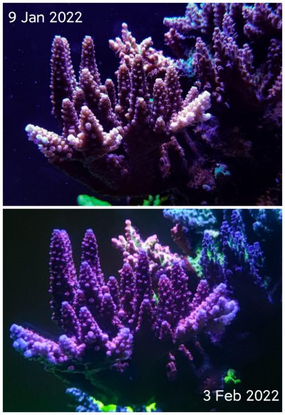
IMG_20220203_225113.jpg

Consider these two photos of the same red stag. They were both taken under full blue light, with a yellow filter for the phone camera. The UV light bar is off for both of the pictures. There is visible increase in the amount of fluorescent proteins on the stag coral. The growth is good, but unquantified.

This is not conclusive even by a far stretch, but the observation is certainly encouraging. Thus I undertake to carry out better experiments to investigate this topic.
 

damsels are not mean

Valuable Member
View Badges
Joined
Nov 14, 2021
Messages
1,952
Reaction score
2,160
Location
Chicago
Rating - 0%
0   0   0
I think to effectively control this you might need more frags and a few different LEDs added. We understand already that fluorescent proteins are related to spectrum, but I think it is not fair to compare adding a spike on the graph in the UV range with broadband increase in intensity. I would suggest adding an equal amount of intensity in the form of other colored LEDs and not just increasing the white.

For example:

-Control with increased broadband PAR
-Experimental group with UV strip
-Experimental group with an equal amount of royal blue added
-Experimental group with red (photo peak) added

It's possible that the addition of a bunch of light at a single wavelength causes the additional production of a particular fluorescent protein that isn't necessarily just a result of increased general intensity but also isn't just something that happens under UV specifically.

This way we can be sure it's actually UV having an effect and not simply the fact that the coral has to adapt to a spike on the chart.

Maybe also include a violet (non UV) LED as another experimental group since LEDs below that wavelength get rapidly more expensive and shorter lived.

Ideally we'd also test this on many corals from different parts of the reef but that's for another day.
 
OP
OP
J1a

J1a

Well-Known Member
View Badges
Joined
Nov 14, 2021
Messages
666
Reaction score
950
Location
Singapore
Rating - 0%
0   0   0
I think to effectively control this you might need more frags and a few different LEDs added. We understand already that fluorescent proteins are related to spectrum, but I think it is not fair to compare adding a spike on the graph in the UV range with broadband increase in intensity. I would suggest adding an equal amount of intensity in the form of other colored LEDs and not just increasing the white.
Thank you for the suggestions.

More frags I can do, but if gonna go for different color led, the whole experiment will be too complex. The added light intensity will not be white. I just plan to raise the frag to increase par without much change in spectrum.

Maybe also include a violet (non UV) LED as another experimental group since LEDs below that wavelength get rapidly more expensive and shorter lived.
There is plenty of violet diodes in many light set. Will increase violet improve the outcome? Maybe. For this experiment though, I want to focus first on what the light sets don't have at all.

And yes. $$ and durability is the likely the reason why we are missing out.
 
OP
OP
J1a

J1a

Well-Known Member
View Badges
Joined
Nov 14, 2021
Messages
666
Reaction score
950
Location
Singapore
Rating - 0%
0   0   0
One more reason I decide to take this approach is that, if the UV indeed is helpful, the easiest way to implement it is through a supplementary light set. Rather than replace the entire display light.

Hopefully a small cost for a big reward.
 

taricha

5000 Club Member
View Badges
Joined
May 22, 2016
Messages
6,970
Reaction score
10,747
Rating - 0%
0   0   0
One more reason I decide to take this approach is that, if the UV indeed is helpful, the easiest way to implement it is through a supplementary light set. Rather than replace the entire display light.
Agree. I think this is a well-formed experiment to answer a narrow practical question. It doesn't have to answer every related question to be helpful. (Though, if a benefit is established - could you get the same benefit with violet lighting as with UV might be a worthwhile follow-up)

Oh, I don't think we said it anywhere that I saw - but obviously the frags will need to be under the same lighting for the photography. Not obvious what lighting that should be, but the control lighting seems a logical choice. If UV changes the color appearence when photographed even under non-UV lighting - that would be a more clear result / confirmation.
 
OP
OP
J1a

J1a

Well-Known Member
View Badges
Joined
Nov 14, 2021
Messages
666
Reaction score
950
Location
Singapore
Rating - 0%
0   0   0
Oh, I don't think we said it anywhere that I saw - but obviously the frags will need to be under the same lighting for the photography. Not obvious what lighting that should be, but the control lighting seems a logical choice. If UV changes the color appearence when photographed even under non-UV lighting - that would be a more clear result / confirmation.
Definately, same control lighting for the photos.

In fact, the photos I attached in post #8 are taken under the same light. For the experiment, I will take the photo of the frags in the same frame, as well as individually, under the same light.
 

MnFish1

10K Club member
View Badges
Joined
Dec 28, 2016
Messages
24,326
Reaction score
23,111
Location
Midwest
Rating - 100%
1   0   0
Don't know if you started this yet - am I understanding the experiment is 1 week? or is it 1 week to ramp up the light? (Maybe I missed it - I didn't see the total duration). If it is a longer duration - I would have some plan about measuring Alkalinity, Ca, etc - in case one side takes off and something starts to be depleted in the whole tank. This will be very interesting. I hope you will keep it up for a longer duration
 
OP
OP
J1a

J1a

Well-Known Member
View Badges
Joined
Nov 14, 2021
Messages
666
Reaction score
950
Location
Singapore
Rating - 0%
0   0   0
Don't know if you started this yet - am I understanding the experiment is 1 week? or is it 1 week to ramp up the light? (Maybe I missed it - I didn't see the total duration). If it is a longer duration - I would have some plan about measuring Alkalinity, Ca, etc - in case one side takes off and something starts to be depleted in the whole tank. This will be very interesting. I hope you will keep it up for a longer duration
It's a one week ramp up. how long the experiment will be really will depends on the result along the way. If there is no difference between the experiment and control groups after 2 month, then I will consider the result negative. If there are differences, I would continue as long as change is observed.

I'm doing it with an LFS, most likely will be using a section of their frag tank. The parameters should be monitored and maintained. But I will do a weekly recording as well.
 

MnFish1

10K Club member
View Badges
Joined
Dec 28, 2016
Messages
24,326
Reaction score
23,111
Location
Midwest
Rating - 100%
1   0   0
It's a one week ramp up. how long the experiment will be really will depends on the result along the way. If there is no difference between the experiment and control groups after 2 month, then I will consider the result negative. If there are differences, I would continue as long as change is observed.

I'm doing it with an LFS, most likely will be using a section of their frag tank. The parameters should be monitored and maintained. But I will do a weekly recording as well.
if it was in 2 different tanks - it would be a 'bigger' potential problem - but the way you're doing it - both sides has the same water - very smart IMHO
 
OP
OP
J1a

J1a

Well-Known Member
View Badges
Joined
Nov 14, 2021
Messages
666
Reaction score
950
Location
Singapore
Rating - 0%
0   0   0
if it was in 2 different tanks - it would be a 'bigger' potential problem - but the way you're doing it - both sides has the same water - very smart IMHO
Thank you! I'm trying to keep as much factors unchanged as possible. It's also fortunate that the lfs owner is keen on doing this. If not, I would be hard-pressed to find space to set up a new tank to do this.

I'll be going to the shop this Friday to work out the details of the set up. I will report on the detailed methodology after that.
 

Dennis Cartier

Valuable Member
View Badges
Joined
Aug 25, 2016
Messages
1,970
Reaction score
2,409
Location
Brampton, Ontario
Rating - 0%
0   0   0
Great experiment.

Don't discount the 420nm in the OR3 having an impact. I am not sure if your main fixtute already provided ample 420nm or not.

In the past, when blue and white LEDs were the norm, I built my own LED fixtures using mostly COB modules from Ebay. The violet portion of the units were 420nm and 430nm 20W modules. I noticed that my SPS had tendency to grow towards the 420 and 430 modules. That was with a lot more LEDs in the 450, 460 and 470+ in the fixture than the few 420 and 430 ones.

I expect you will see an effect.

Dennis
 
OP
OP
J1a

J1a

Well-Known Member
View Badges
Joined
Nov 14, 2021
Messages
666
Reaction score
950
Location
Singapore
Rating - 0%
0   0   0
Great experiment.

Don't discount the 420nm in the OR3 having an impact. I am not sure if your main fixtute already provided ample 420nm or not.

In the past, when blue and white LEDs were the norm, I built my own LED fixtures using mostly COB modules from Ebay. The violet portion of the units were 420nm and 430nm 20W modules. I noticed that my SPS had tendency to grow towards the 420 and 430 modules. That was with a lot more LEDs in the 450, 460 and 470+ in the fixture than the few 420 and 430 ones.

I expect you will see an effect.

Dennis
You are right! My original light, while have quite a bit of 420nm, the most intense peak is at 450nm. IMG_20211231_144752.jpg

For the experiment though. I will supplement with UV only for a start, leaving out the 420nm.

Then we can address the 420nm subsequently.
 
OP
OP
J1a

J1a

Well-Known Member
View Badges
Joined
Nov 14, 2021
Messages
666
Reaction score
950
Location
Singapore
Rating - 0%
0   0   0
Physical Setup

After considering the feedback given, I would outline the latest methodology for this investigation.

The investigation will be carried out in a shallow holding tank in a Reefing Reality, a Singapore LFS. They also kindly provided the frags for this project.

Baseline

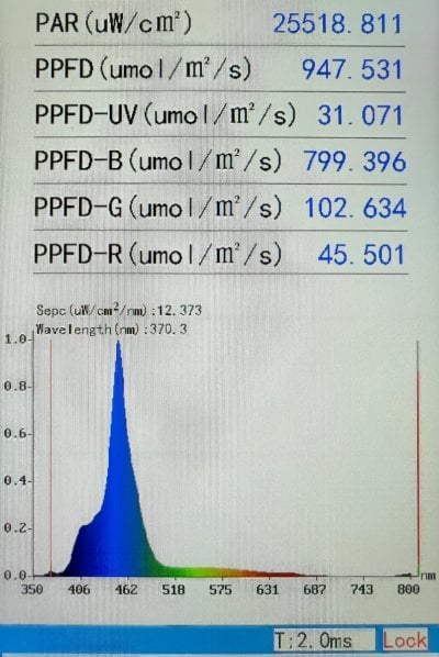
The holding tank is part of the linked SPS holding system, and illuminated by multiple Wyatt lights. The basedline PAR value and spectrum at the reserved experiment sites is shown below.

IMG_20220218_155214.jpg


The baseline PAR is moderate, below photo saturation levels. This allows us some buffer for PAR increase. The spectrum output of the light is characteristic of most modern LEDs. High in 450nm royal blue segments, while providing little in the way of UV, as well as longer wavelengths.


UV Supplement

To supplement UV spectrum for this investigation, we deploy a simple UV ribbon LED. The two 50cm length ribbons has a combined power draw of 22 watt, they are fixed on a parabolic reflector without any refractive optics. These ribbons consist of 365nm and 380nm diodes in 3:1 ratio. The photograph below shows the supplement light set mounted in place.

PSX_20220218_125109.jpg


The spectrum and PAR is shown in the graph below.

IMG_20220218_124514.jpg

Take note of the low but distinct peak observed at 365nm, as compared to the baseline illumination. It also increase the overall PAR.


PAR Matching

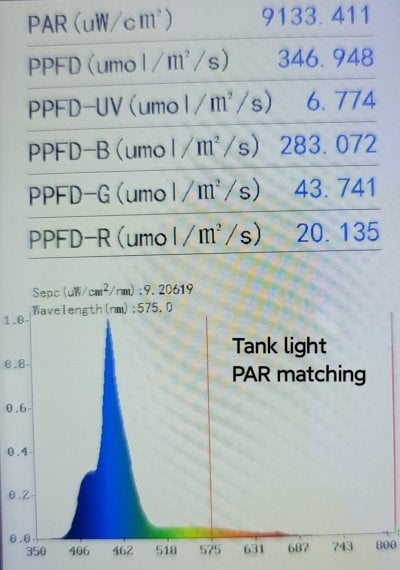
in order to factor in the increased in PAR of the experiment group, a third groups is set up, in the same tank, at a region of higher PAR. This raises the PAR without introducing any additional UV spectrum. The location has a PAR measurements as shown. We will find tune the position to match the PAR value as close as possible once the experiment is underway, and the frags are transferred to this new location.

IMG_20220218_124642.jpg
 

Attachments

  • IMG_20220218_124642.jpg
    IMG_20220218_124642.jpg
    164.2 KB · Views: 54
Back
Top